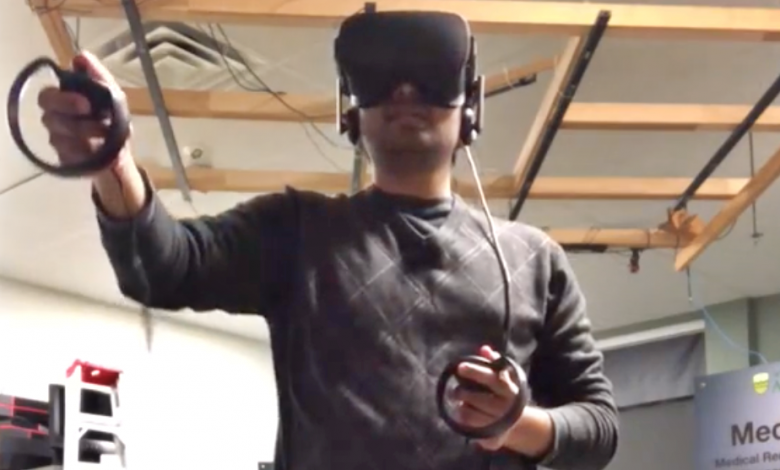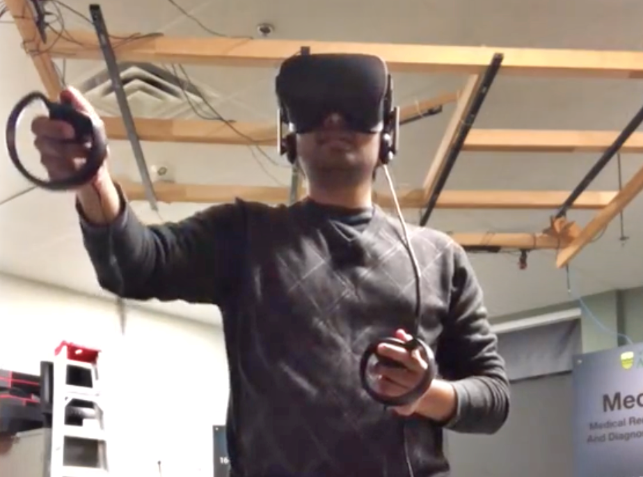
Now that we’ve seen and heard Pierre and Kumar share insights in Episode 1 and 2 about how surgery will advance globally in the near future, we are pleased to give you a sneak preview of the VR Surgical System App.
Chirag Balakrishna and David Cocom Basto play the roles of clinical team participants (host & participants).

To start the host invites the other participants to connect directly into the virtual reality room. All this connection needs is for the participants is an IP and a port set up before running the VR Surgical System app. Once the connection is made, the other participants appear in the room and wave at each other.

Note that both the hands and head actions of the two participants in this demo are tracked in real time.
The dataset in this demo uses a 3D CT scan image of a chest – the ribs, heart, and lungs, which are visible for the participants to grab, rotate and position the object, and to point out a specific part of the 3D CT scan image to other participants as part of the process in surgical planning.
Participants can then slice the 3D CT scan image (object) using the clipping plane attached to the object. This plane allows the participants to slice the object at any angle – or by using each of the available axis (x, y, z) from top to bottom or vice-versa using the menu and the left joy stick.
In the video demo, you’ll see Chirag make annotations and drawings into the object – visible to all participants. Draw options shown in the menu can be used to create annotations in different colours, or to clear the scene.
A scientific article also appears in the VR room demo – a key feature that allows doctors, radiologists and surgical teams to know more about the patient file and / or about specific relevant illnesses or conditions.
You can also make annotations in the article (shown as circles around the words) and point out certain specific terms or highlight key information to relevant about the patient to the participants.
To close the virtual room and end the session, the participants wave to each other to sign off!
Congratulations – you have just participated in the VR Surgical System from your location globally!
Stay tuned for future stages of development by Pierre and Kumar on the VR Surgical System.
About Dr. Pierre Boulanger
Pierre is currently the Director of the Advanced Man Machine Interface Laboratory (AMMI) as well as the Scientific Director of the SERVIER Virtual Cardiac Centre in the Mazankowski Heart Institute. In 2013, Dr. Boulanger was awarded the CISCO chair in healthcare solutions, a 10 years investment by CISCO systems in the development of new IT technologies for healthcare in Canada.
Dr. Boulanger received his in Engineering Physics and his Masters in Physics Laval University, and his Ph.D. in Electrical Engineering from the University of Montreal. Dr. Boulanger cumulates more than 35 years of experience in 3D computer vision, rapid product development, and the applications of virtual reality systems to medicine and industrial manufacturing. Dr. Boulanger worked for 18 years at the National Research Council of Canada as a senior research officer where his primary research interest was in 3D computer vision, rapid product development, and virtualized reality systems. He now has a double appointment as a professor at the University of Alberta Department of Computing Science and at the Department of Radiology and Diagnostic Imaging.
About Dr. Kumaradevan Punithakumar
Kumar is currently the Operational and Computational Director of the SERVIER Virtual Cardiac Centre in the Maznakowski Heart Institute. Dr. Kumaradevan Punithakumar received the B.Sc.Eng. (with First class Hons.) degree in electronic and telecommunication engineering from the University of Moratuwa and the M.A.Sc and Ph.D. degrees in electrical and computer engineering from McMaster University. From 2008 to 2012, he was an Imaging Research Scientist at GE Healthcare, Canada. He was the recipient of the Industrial Research and Development Fellowship by the National Sciences and Engineering Research Council of Canada in 2008, and the GE Innovation award in 2009. Areas of interest include medical image analysis and visualization, information fusion, object tracking, and nonlinear filtering.
About Dr. Michelle Noga
Michelle is the Medical Director of the SVCC. Dr. Michelle Noga is a radiologist trained at University of Alberta and British Columbia’s Children’s and Women’s hospital. She is currently an associate professor at the University of Alberta, department of radiology and diagnostic imaging, and a radiologist with Medical Imaging Consultants. Areas of interest include pediatric cardiac MRI and CT, post-processing of cross-sectional cardiac imaging, 3D visualization, rapid prototyping, pediatric airway, and finite element analysis.
About Student Chirag Balakrishna
Chirag is a second year M.Sc. student in Computing Science at the University of Alberta and worked on the Virtual Reality based Collaborative Surgical Planning application as a Graduate Research Assistant at the Advanced Man Machine Interface Lab (AMMI). He is interested in computer graphics and its applications in the field of scientific visualization such as medical imaging in virtual reality in order to help improve patient care. Chirag graduated with a bachelor in Computer Science and Engineering from PES Institute of Technology, Bangalore. He interned as a Web Developer for Reverie Language Technologies.
About Student David Cocom Basto
David is pursuing a M.Sc. in Computer Science in specializing in Image Processing, Virtual Reality, Graphics and Animation, and Multimedia Communications at the University of Alberta. He worked on the Virtual Reality based Collaborative Surgical Planning application at the SVCC. David has a BSc in Software Engineering, related to the software creation process – starting with the requirements specification, continuing with the design and build, and finishing with the release and maintenance, with consideration from both developer and project manager perspectives.


